5 Draw a Basic Diagram of Spectroscopic Measurements
In the broadest sense a spectrometer is whatever instrument that is used to measure out the variation of a physical feature over a given range; i.eastward. a spectrum. This could be a mass-to-charge ratio spectrum in the example of a mass spectrometer, the variation of nuclear resonant frequencies in an NMR spectrometer or the change in the absorption and emission of light with wavelength in an optical spectrometer.
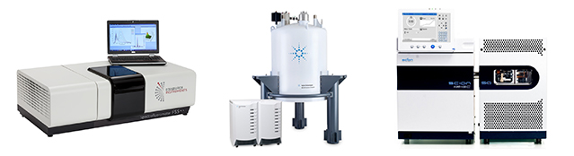
Effigy 1: The three nearly common types of spectrometers found in research labs around the world.
Left: Optical spectrometer (Edinburgh Instruments FS5 spectrofluorometer).
Centre: NMR spectrometer (Agilent 800 MHz NMR spectrometer).
Right: Mass spectrometer (Scion Instruments GC-MS spectrometer).
The about ubiquitous type of spectrometer used for research are optical spectrometers; and when someone simply says 'spectrometer', without an boosted qualifier, they are normally referring to an optical spectrometer and this diverse family unit of spectrometers is the focus of this article.
How does an Optical Spectrometer work?
The goal of any optical spectrometer is to measure out the interaction (absorption, reflection, scattering) of electromagnetic radiation with a sample or the emission (fluorescence, phosphorescence, electroluminescence) of electromagnetic radiation from a sample. Optical spectrometers are concerned with electromagnetic radiation that falls within the optical region of the electromagnetic spectrum which is light spanning the ultraviolet, visible and infrared wavelength regions of the spectrum.
In order to gain maximum information, the interaction or emission of light should be measured as a function of wavelength and the common feature of all optical spectrometers is therefore a mechanism for wavelength option. In low price spectrometers or in situations where accurate wavelength selection is non important, optical filters are used to isolate the wavelength region of interest. However, for authentic wavelength selection and the generation of spectra, a dispersive element that separates light into its elective wavelengths is required. In all modern spectrometers, this dispersive element is a diffraction grating where constructive and destructive interference is used to spatially separate polychromatic lite that is incident on the grating (Figure 2).
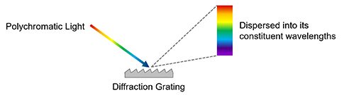
Figure 2: Dispersion of low-cal into its constituent wavelengths by a diffraction grating.
Diffraction gratings are a primal component of a monochromator, which is a device used to select a particular wavelength of low-cal from a polychromatic light source. In a monochromator the diffraction grating is rotated to change the wavelength that aligns with and passes through the exit slit. Excitation monochromators are institute in all spectrophotometers (come across following department) for selecting the desired excitation wavelength to reach the sample from a white lite source (Figure 3 Excitation Monochromator) and spectra are measured past scanning the monochromator and measuring the modify in signal as a role of excitation wavelength.
For detecting the low-cal emitted by a sample at that place are two approaches. The start is an emission monochromator which works using the same principle equally above except the low-cal source is the emission from a sample and the monochromator selects which wavelength of low-cal reaches the detector (Figure iii Emission Monochromator). The 2d approach is to detect the spectrum of the dispersed light 'all at once' using an array detector (such as a CCD camera) which is called a spectrograph (Figure 3 Spectrograph). At least one emission monochromator or spectrograph is found in all spectrofluorometers and Raman spectrometers (see post-obit sections).
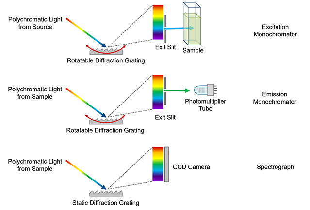
Figure three: The bones operating principle behind monochromators and spectrographs. It should be noted that these images are highly simplified for analogy. For example the monochromators used in the Edinburgh Instruments FS5 and FLS1000 are a more complicated Czerny-Turner blueprint which take two slits and 2 ellipsoidal mirrors for superior performance simply the principle remains the same.
Types of Optical Spectrometer
At present that the key component of a spectrometer has been identified, the different types of spectrometer, their office, and basic pattern tin be discussed. Three of the virtually common optical spectrometers: spectrophotometers, spectrofluorometers and Raman spectrometers are introduced.
Spectrophotometer (also known as a UV-Vis Spectrometer)
The term spectrophotometer can refer to quite a variety of instruments that measure light, with the exact definition depending on the area of science or manufacture. In all cases the term 'photo' is used to indicate that the spectrometer is for the quantitative measurement of calorie-free intensity with wavelength. Inside academic inquiry (particularly chemistry and biology laboratories) the term spectrophotometer is used specifically to refer to a spectrometer which measures the absorption of low-cal past a sample and that definition will be used here.
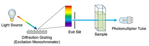
Effigy iv: Simplified diagram of a single beam spectrophotometer.
A stylised layout of a basic unmarried beam spectrophotometer is shown in Effigy 4. Information technology comprises a white light source which is usually either a combination of a deuterium arc lamp to cover the UV range and a tungsten halogen lamp to cover the visible; or a single Xenon arc lamp to cover the unabridged range. This is followed by an excitation monochromator which selects the wavelength of calorie-free that reaches the sample. This lite is and so either transmitted through the sample (equally shown in Figure 4) for transparent samples such as solutions or reflected off the surface for opaque samples. The intensity of the transmitted or reflected lite is then monitored using a detector which is typically a photomultiplier tube or silicon photodiode.
In addition to stand up alone spectrophotometers, the functionality of a spectrophotometer tin can also be incorporated as a secondary characteristic into other spectrometers. An instance of this is the FS5 Spectrofluorometer which comes with a transmission detector equally standard and has all the functionality of a unmarried beam spectrophotometer in add-on to existence a spectrofluorometer.
The nearly mutual measurement undertaken in a spectrophotometer is measuring the absorption spectrum of a sample. The excitation monochromator is scanned and the change in calorie-free intensity transmitted through the sample recorded on the detector. This is then repeated with a reference sample and the absorption spectrum calculated equally shown in Figure 5 for a solution of fluorescein in phosphate buffered saline.
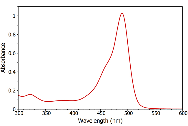
Effigy v: Absorption spectrum of fluorescein measured using the FS5 Spectrofluorometer.
A much more advanced version of a spectrophotometer is a transient absorption spectrometer, which can measure the development of the assimilation spectrum with time and is essential for looking at temporary species generated through chemical reactions or short lived photoexcited states. The performance and design of transient absorption spectrometers are beyond the scope of this commodity but you can learn more in our introduction to transient absorption using the Edinburgh Instruments LP980 Transient Absorption Spectrometer.
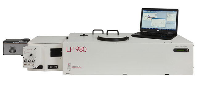
Figure 6: Edinburgh Instruments LP980 Transient Assimilation Spectrometer.
Spectrofluorometer (besides known equally Fluorescence/ Photoluminescence Spectrometer)
A spectrofluorometer is used to measure the fluorescence emission (or more generally, the photoluminescence) from a sample. The terms spectrofluorometer and fluorescence / photoluminescence spectrometer are interchangeable and different manufacturers telephone call them past different names. A general convention is that spectrofluorometer refers to a meaty benchtop musical instrument that is similar in size to a spectrophotometer, such as the FS5 Spectrofluorometer, while the term fluorescence / photoluminescence spectrometer is used for larger spectrometers with superior operation and more than diverse functionality such as the FLS1000 Photoluminescence Spectrometer.
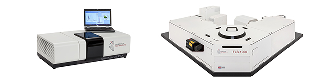
Figure 7: Example of a compact spectrofluorometer (FS5) and a more advanced photoluminescence spectrometer (FLS1000).
The layout of a typical steady state spectrofluorometer is shown in Figure 8. The excitation side of a spectrofluorometer is equivalent to the spectrophotometer: a white light source and an excitation monochromator. Xenon arc lamps are used every bit the calorie-free source every bit their loftier brightness is essential to measure the weak fluorescence emission. The sample is illuminated by the chosen excitation wavelength which causes information technology to fluoresce. The fluorescence emission is collected by the emission monochromator which is orientated at xc degrees to the excitation monochromator and the selected wavelength reaches the detector; typically a photomultiplier tube. For more detail on the blueprint of spectrofluorometers read our article on fluorescence instrumentation.
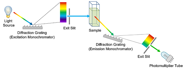
Figure 8: Simplified diagram of a spectrofluorometer.
The two most common spectral measurements undertaken in a spectrofluorometer are excitation and emission spectra. To measure an excitation spectrum the emission monochromator is ready at a wavelength of strong fluorescence emission, the excitation monochromator is scanned beyond the wavelength region of involvement and the change in fluorescence intensity monitored on the detector (Effigy 9 blue curve) which reveals the absorption features of sample. Emission spectra are the inverse of this; the wavelength of the excitation monochromator is set where in that location is strong absorption by the sample, the emission monochromator is swept across the wavelength region of interest and the alter in fluorescence detected (Figure ix red curve) revealing the fluorescence features of the sample.
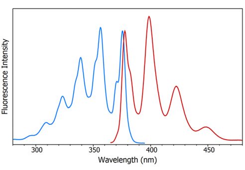
Figure 9: Fluorescence excitation (blue) and emission (red) spectra of anthracene measured using the FS5 Spectrofluorometer.
Raman Spectrometer
Raman spectrometers are used to mensurate the Raman scattering of lite from a sample. The design of a typical Raman spectrometer is shown in Figure ten and is similar to a spectrofluorometer but with a few cardinal differences. The white light source and excitation monochromator found in spectrofluorometers are replaced with a laser. The reason for this is twofold. The starting time is that 'Raman' is a scattering issue and the lite is therefore not absorbed by the sample. This ways that a broadband tuneable calorie-free source for matching to the absorption features is not required. The 2d reason is that the Raman outcome is much weaker than fluorescence (ratio of Rayleigh scattered low-cal to Raman scattered low-cal is ~ten6) and sources with a high photon flux are therefore essential to maximise the indicate.
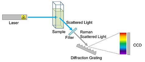
Effigy 10: Simplified diagram of a Raman spectrometer.
The laser light is scattered off the sample and passed through a filter to remove the much more than intense Rayleigh scattered component. The remaining Raman scattered calorie-free is and so passed into a spectrograph and captured using a CCD detector to give a Raman spectrum like that shown in Effigy 11 of paracetamol.
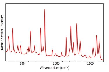
Figure xi: Raman spectrum of paracetamol.
Fluorescence Spectrometer Instrumentation
We hope this article on the types of spectrometer has provided a useful introduction to the most ordinarily encountered optical spectrometers and the type of measurements that they can produce. For more than advanced information on fluorescence spectroscopy and spectrometers why not read our web log or explore our range of fluorescence and transient absorption spectrometers.
Keep in Touch
If yous take enjoyed reading this blog mail, and want to be the first to see all the latest news, applications and product information, then sign-upwards to our infrequent newsletter via the red sign-up push button below and join us on social media.
5 Draw a Basic Diagram of Spectroscopic Measurements
Source: https://www.edinst.com/blog/what-is-a-spectrometer/
Post a Comment for "5 Draw a Basic Diagram of Spectroscopic Measurements"